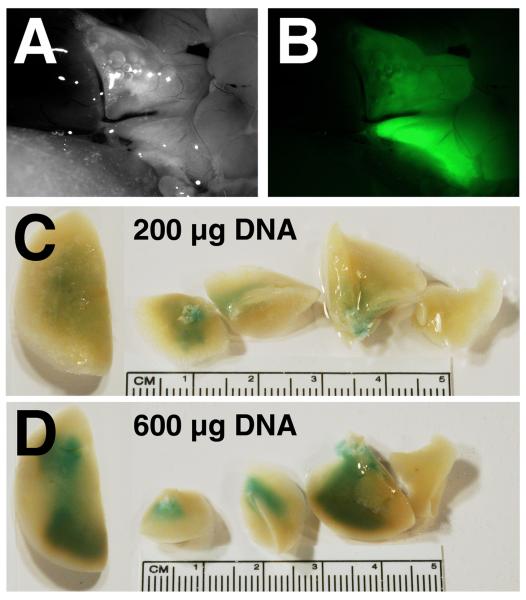
Figure 2. Distribution of gene delivery and expression in electroporated rat lungs.
A and B. GFP-β1 expressing plasmids (600 μg) were administered to the lungs and electroporated (200 V/cm, 8 pulses at 10 μsec each). Three days later, the lungs were visualized in situ (A) and GFP-β1 expression was detected by fluorescence microscopy (B). C and D. Dose-dependency of gene transfer and expression. Two hundred micrograms (C) or 600 μg (D) of a β-galactosidase-expressing plasmid was transferred to lungs and electroporated (200 V/cm, 8 pulses at 10 μsec each). Three days later, the lungs were removed and reacted with X-gal to visualize β-galactosidase expression.

