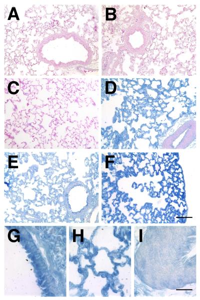
Figure 3. Localization of gene expression in electroporated lungs.
β-galactosidase-expressing plasmids were transferred to rat lungs (n=6), electroporated (200 V/cm, 8 pulses at 10 μsec each), and three days later, the lungs were removed, inflated to total lung capacity, fixed, and paraffin-embedded for sections. Immunohistochemistry was performed with antibodies against β-galactosidase using the Vector-Blue ABC reagent and sections were counterstained with eosin (A – F). A. Naïve lung. B. Electroporation only (no plasmid). C. DNA only (no electroporaiton). D – I. Plasmid with electroporation. At high magnification, airway epithelial and smooth muscle cells (G), alveolar type I and type II cells (H), and vascular smooth muscle cells (I) can be seen to express transgene. A – F, Bar = 100 μm; G – I, Bar = 20 μm.

