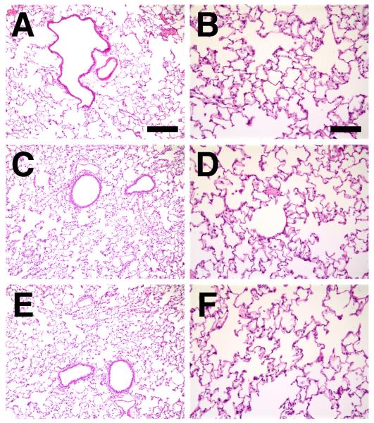
Figure 4. Histological analysis of electroporated lungs.
Lungs from naive animals (A), or those that were electroporated (100 V/cm, 8 pulses at 10 msec each) without added DNA (C and D) or with 600 μg of pcDNA3 (empty vector; E and F) were harvested three days post-electroporation, inflated to total lung capacity, fixed, paraffin-embedded, sectioned and stained with hematoxylin and eosin. Panels A, C, and E were taken at the same magnification (bar = 200 μm), and panels B, D, and F were taken at a higher magnification (bar = 100 μm). Representative sections from one of 3 animals at each condition are shown.

