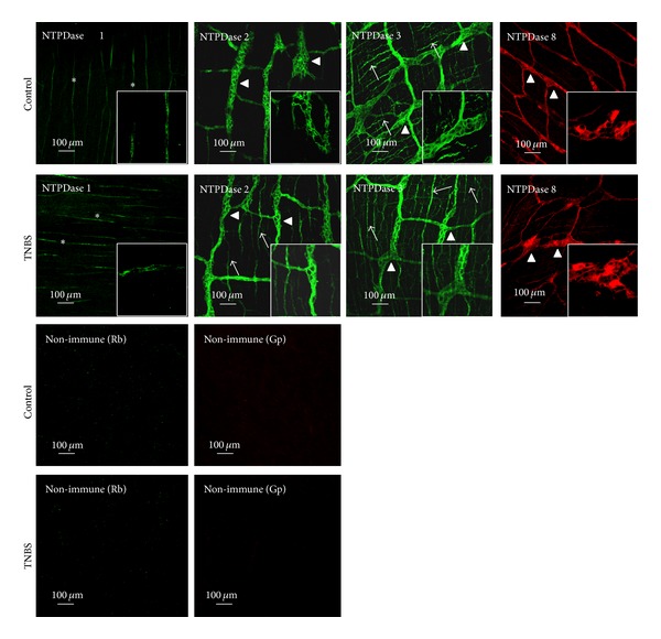
Figure 8.

Localization of NTPDase1, NTPDase2, NTPDase3, and NTPDase8 immunoreactivity in single confocal images of whole-mount preparations of the longitudinal muscle-myenteric plexus of the ileum of control and TNBS-injected rats. In healthy animals, NTPDase 1 immunoreactivity is present only in blood vessels (asterisks); NTPDase2 immunoreactivity is present predominantly in ganglion neuronal cell bodies (triangles) and large ramifications (primary meshwork) of the myenteric plexus, whereas NTPDase3 is also evident on myenteric axon terminals (arrows). In TNBS-treated preparations the NTPDase2 staining acquires a pattern that is very similar to that of NTPDase3, with NTPDase2 immunoreactivity also appearing in nerve bundles and axon terminals of myenteric neurons. NTPDase8 stains few ganglion neuronal cell bodies and large ramifications (primary meshwork) of the myenteric plexus; this pattern did not significantly change among control and TNBS-treated animals. No staining was obtained when nonimmune sera from host species (rabbit, Rb, and guinea-pig, Gp) were used instead of interest primary antibodies.
