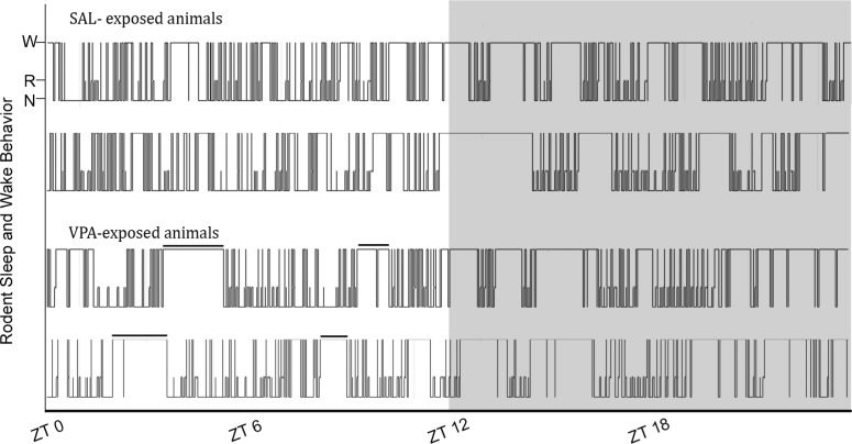
Figure 1.
Representative hypnograms of juvenile rats exposed to saline (SAL, top) and valproic acid (VPA, bottom) in utero across a 24-h period. W, wake; N, NREM sleep; R, REM sleep. The solid black bar is highlighting consolidated wake bouts during the light phase of VPA-exposed rats, which are absent in the SAL-exposed controls. Gray area is lights off.

