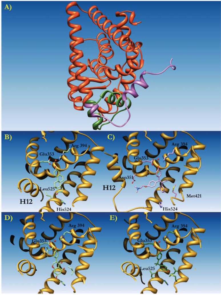
Fig. (4).
Molecular Modeling of the estrogen receptor ERα complexed to diverse ligands. A) The three dimensional structure of human ERa represented as orange ribbons. Helix XII is colored in green when positioned in the active conformation, and in purple when in an inactivated (i.e. protein bound to an antagonist such as Tamoxifen) conformation. B) Detail of the conformation of Helix XII (H12) with the natural ligand 17-b-estradiol (E2, light green) bound to the active site. Residues involved in ligand binding are drawn as sticks. The active water molecule is represented by a red sphere. C) The conformation of Helix XII (H12) when binding hydroxytamoxifen. D) The conformation of Helix XII (H12) when binding Lasofoxifene. E) The conformation of Helix XII (H12) when binding Bisphenol A.

