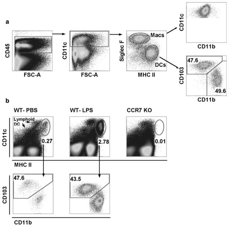
Fig. 1.

Gating strategy for the identification of DCs in lung and LLN. a Whole lung digest from a naïve mouse gated on live cells. CD45+ cells were plotted for CD11c expression. CD11c+ cells were plotted as Siglec-F versus MHC II to differentiate macrophages from DCs. Gated Siglec-F− MHCII+ DCs were plotted as CD103 versus CD11b to differentiate migratory DC subsets that are found in similar frequencies. b Live cells from single LLNs 24 h post-intranasal instillation of PBS or 2 μg LPS isolated from WT or CCR7−/− mice. Cells plotted as CD11c versus MHC II display lymphoid-resident and migratory DC populations. Migratory DCs have higher MHC II expression than lymphoid-resident DCs. CD11c+ MHCIIhi migratory DCs were plotted CD103 versus CD11b to demonstrate similar migration frequencies in WT mice
