
Figure 2. Ureaplasma urealyticum ascends into the uterus in MHV68 infected animals.
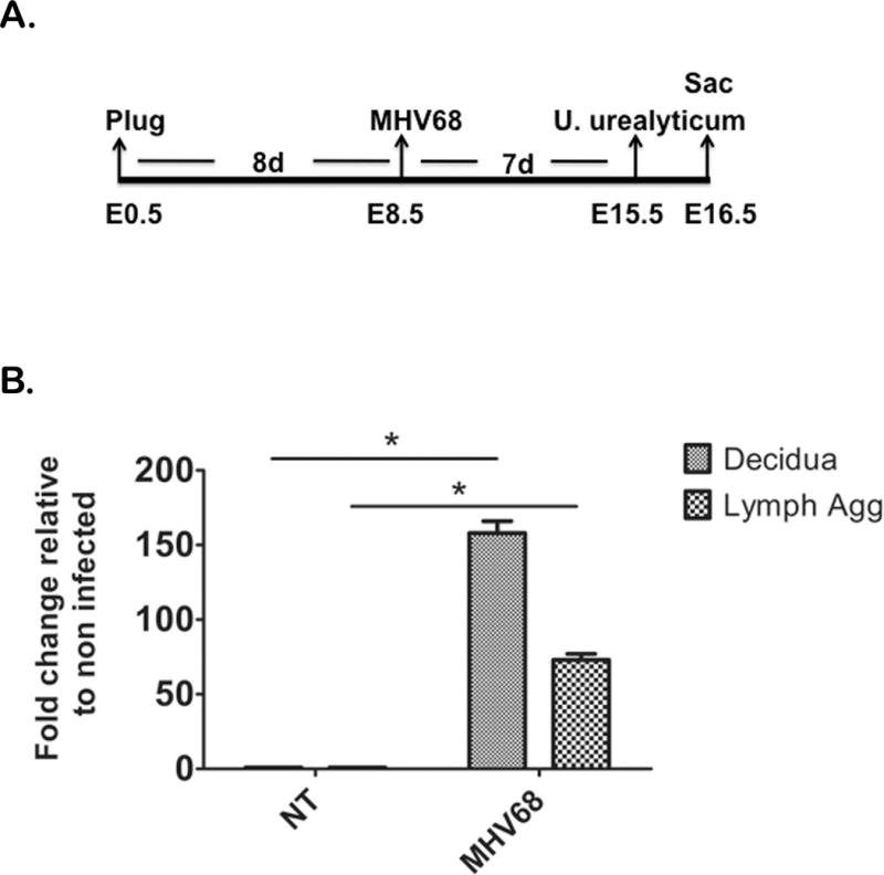
Mice were infected with MHV68 or DMEM (control) at E8.5 of pregnancy, received an intravaginal injection of pathogenic U. urealyticum at E15.5, and were sacrificed (Sac) at E16.5 (A). NT= mice with no MHV68, MHV68= virus treated animals. Both groups received U. urealyticum; decidua and lymphoid aggregates are represented as fold difference from the same tissue from mice with no viral infection. Mice with MHV68 had higher concentrations of bacteria in the decidua and lymphoid aggregates (Lymph Agg) compared to mice without viral infection (B). n=5