
Fig. 3.
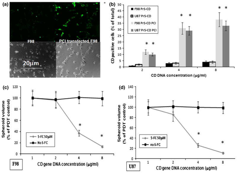
Effects of PCI-CD on gene transfection and on sensitivity of F98 and U87 spheroids to 5-FC. a Fluorescence microscopy images of F98 monolayer with and without PCI transfected CD gene cells. Cell monolayers incubated with anti-CD antibody and a second tagged antibody with green fluorescence; left panel F98 fluorescent and phase contrast, respectively; right panel transfected cells, fluorescent and phase contrast, respectively; green CD gene product labeled with fluorescent antibody. b % of CD positive F98 and U87 cells as determined from fluorescence microscopy images compared to identical regions on phase contrast images. c Effects of 5-FC on the growth and development of F98 and U87 spheroids following PCI-CD gene transfection. Spheroids incubated 18-h with PrS-CD polyplexs at CD DNA concentrations shown in the figure and AlPcS2a. Light treatment, 1.5 J/cm2 @ 5 mw/cm2. 50 μM 5-FC added to groups as shown in figure. Each experimental point represents the results of 24 replicate cultures from 3 independent experiments and is shown as averaged percentages of PDT control cultures ± SE. Asterisks represents significance compared to control values at p <0.05