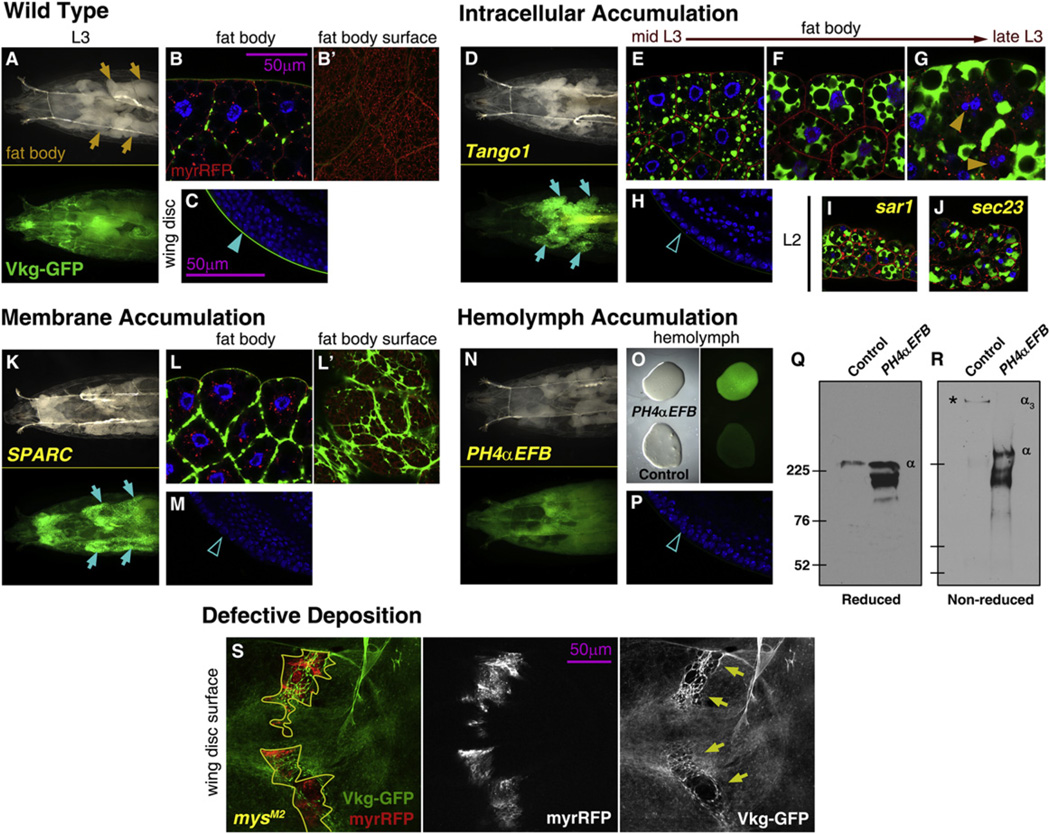
Figure 4. Multiple Requirements for Correct Collagen IV Incorporation into BMs.
(A) Anterior half of a vkgG454/+ L3 larva. Green fluorescence in lower subpanel.
(B) Confocal image of fat body cells of a vkgG454/+ L3 larva. Nuclei in blue (DAPI), Vkg-GFP in green and membranes in red (UAS-myrRFP). B’ shows a surface section.
(C) Confocal image of the wing disc (posterior ventral hinge) of a vkgG454/+ L3 larva. Nuclei in blue (DAPI), Vkg-GFP in green.
(D–H) Images of vkgG454/+ L3 larvae where expression in the fat body of the cargo adaptor Tango1 has been knocked down, showing the anterior half of the larva (D, compare to A), fat body cells (E–G, compare to B) and wing disc (H, compare to C).
(I and J) Confocal images of the fat body of vkgG454/+ L2 larvae where expression of the CopII components Sar1 (I) and Sec23 (J) has been knocked down.
(K–M) Images of vkgG454/+ larvae where SPARC expression in the fat body has been knocked down, showing the anterior half of the larva (K), fat body cells (L, surface in L’), and wing disc (M).
(N–P) Images of vkgG454/+ L3 larvae where PH4-αEFB expression in the fat body has been knocked down, showing the anterior half of the larva (N), bled hemolymph (O, bright-light image on left and green fluorescence on right) and wing disc (P).
(Q and R) Anti-GFP Western blots of hemolymph from control larvae and larvae where PH4αEFB expression in the fat body has been knocked down, both vkgG454/+ (as in O). SDS-PAGE gels were run under reducing conditions (Q) which separate Collagen IV monomers (α) or nonreducing conditions (R), which preserve the Collagen IV trimers (α3). Predicted molecular weight of a Vkg-GFP monomer is 221 kDa (Vkg 194 kDa + GFP 27 kDa).
(S) Clones of mysM2 (integrin βPS) mutant cells in vkgG454/+ wing discs. Clones, outlined in yellow, are labeled by myrRFP expression (red on left and white in middle). Arrows point to scars in the Vkg-GFP layer (green on left and white on right).
See also Figure S3.

