
Figure 4. Cell-associated and soluble antigens are processed differently in CD103+ DCs.
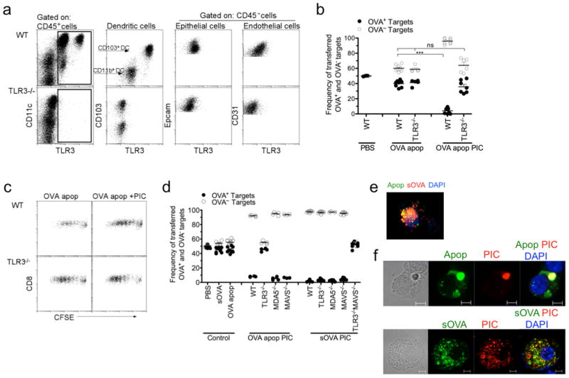
a, TLR3 protein expression of hematopoietic (CD45+) and non-hematopoietic (CD45-) cells isolated from the lungs of WT and TLR3-/- mice. Dendritic cells were identified as low SSC CD11c+MHCIIhi cells prior to being plotted as CD103 versus TLR3. b,d, OT-I CD8 T cells were adoptively transferred into WT, TLR3-/-, MDA5-/-, MAVS-/- or TLR3-/-MAVS-/- mice 1d prior to i.n. delivery of PBS, soluble OVA or OVA+ apoptotic cells +/- 10μg Poly I:C (PIC). Five days after immunization, mice were given PBSE-labeled target (OVA— or OVA+) cells. 24h later spleens were assessed for target cell frequency. Each dot represents one mouse. Data represents at least 3 independent experiments with 3-4 mice per group. ***p< 0.0001 t-test. c, CFSE-labeled OT-I CD8 T cells (1×10⁶ total cells) were adoptively transferred into WT and TLR3-/- mice 1d prior to i.n. delivery of OVA+ apoptotic cells +/- 10 μg Poly I:C (PIC). T cell proliferation was assessed 3d later by FACS. e, Microscopy analysis of isolated pulmonary CD103+ DCs 2 hours after intranasal co-delivery of CFSE-labeled apoptotic cells (green) and soluble OVA-Alexa 647 (red); DAPI (blue). f, Microscopy analysis of isolated pulmonary CD103+ DCs 2 hours after intranasal co-delivery of CFSE-labeled apoptotic cell (green) or soluble OVA-FITC (green) with Rhodamine-labeled Poly I:C (red); DAPI (blue). Scale bar represents 5μm. Data represents at least 4 independent experiments with 10-20 CD103+ antigen-bearing DCs analyzed per slide.