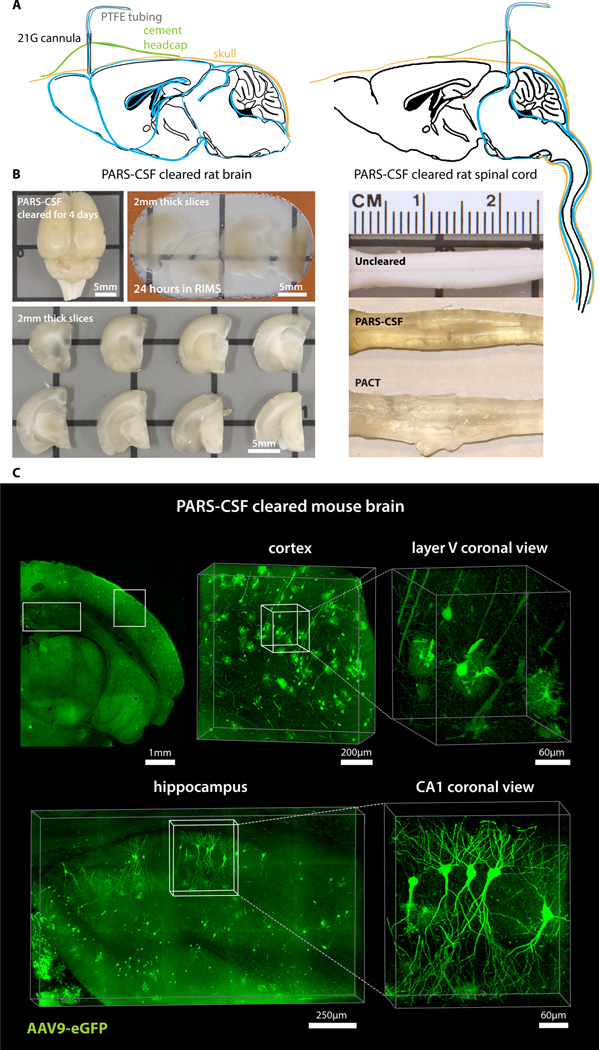
Figure 3. PARS-CSF: a protocol for rapid whole-brain or spinal cord clearing and labeling via the cerebrospinal fluid route (CSF) using perfusion-assisted agent release in situ (PARS).
(A) CNS tissue may be rendered transparent optically transparent by the direct perfusion of all PARS reagents into the CSF via an intracranial brain shunt inserted either (left) below the dura in the region directly above the olfactory bulb, or into the cisterna magna (or placed directly above the dorsal inferior colliculus, right). The cannula, which is connected to the perfusion lines may be cemented into position with dental acrylic. (B) Whole-brain and the corresponding 2 mm thick slices (left) and whole-spinal cord (right) from PARS-CSF rats that were cleared at 37 °C for 4-days (brain) or for 2-weeks (spinal cord) are shown. The extent of whole-brain clearing is dependent on brain tissue proximity to the cannula: the frontal lobe was rendered optically transparent, whereas the mid-hind brain were only weakly cleared (see 2 mm slices on right side of panel). After 24-hour incubation in RIMS, PARS-CSF brain slices were sufficiently cleared for imaging without further sectioning. C) Images show native eGFP fluorescence in 500 µm PARS-CSF cleared coronal brain slices prepared from mice that, 6-months prior to clearing, received IV injections with AAV9:CAG-eGFP. Representative sections of cortex and hippocampus are presented at higher magnification in image boxes (right). In the layer V coronal view, an AAV9 transduced eGFP-expressing glial cell and eGFP-neuron adjacent to a blood vessel are clearly visible. In the hippocampus (bottom), the finer neuronal processes of eGFP-expressing CA1 neurons may be visualized with high resolution, which suggests that PARS-CSF may be completed without severe damage to cellular morphology. For microscopy see Supplemental Methods. Also see Figure S4.

