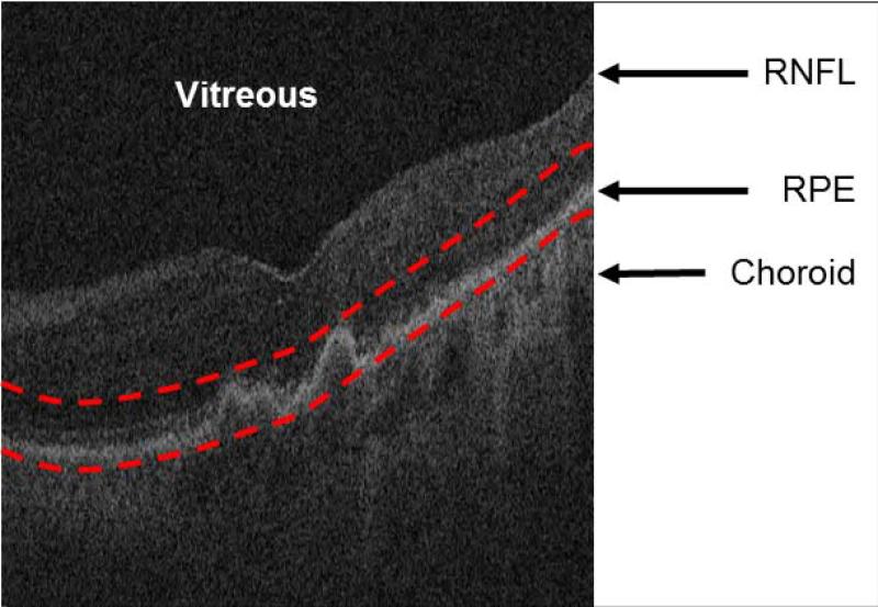
Figure 2.
Restricting the optical coherence tomography image data to the vicinity of the retinal pigment epithelium (RPE) layer for generating the restricted summed-voxel projection (RSVP). The bottom red curve is the baseline of the normal RPE layer. The top red curve is determined by the largest drusen height in all of B-scans. The RSVP thus excludes extraneous portions of the retina that may contain noise to the projection, such as those caused by the vitreous, retinal nerve fiber layer (RNFL), and choroid.

