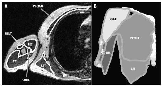
Figure 1.

Three-dimensional (3D) muscle surfaces created from axial MR images. (A) Example axial slice showing the boundaries of the shoulder adductors (medium gray), shoulder abductors (white), elbow flexors (dark gray), and elbow extensors (light gray). In this slice, visible shoulder adductors include pectoralis major (PECMAJ), latissimus dorsi (LAT), and coracobrachialis (CORB); visible abductors include deltoid (DELT); visible elbow flexors include biceps (BIC); and the visible elbow extensor is triceps (TRI). (B) A 3D surface rendering of each muscle is created from the segmented images to determine muscle volume.
