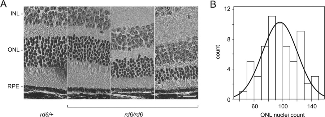
Fig. 1.
Phenotypic analysis of retinas at 20 weeks of age. (A) ONL thickness of F2 homozygous Mfrprd6 mice varies in a segregating genetic background obtained from a (B6.C3Ga- Mfrprd6/J × CAST/EiJ) F1 intercross. Heterozygous mice (rd6/+, left panel) show normal retinal layers while homozygous Mfrprd6 progeny (rd6/rd6) showed differing degrees of ONL thinning. The Mfrp genotype is indicated. INL, inner nuclear layer; ONL, outer nuclear layer; RPE, retinal pigment epithelium. Bar: 20 µm. (B) Frequency distribution of ONL nuclear counts from 63 F2 homozygous Mfrprd6 mice. The data fit a normal distribution (solid curve).

