
Figure 1.
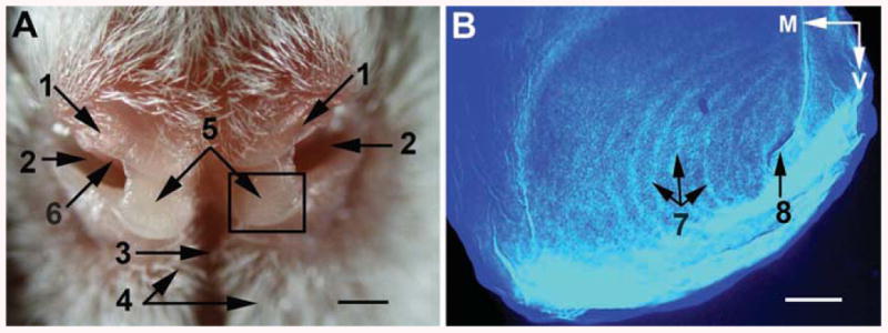
Morphology of the rat rhinarium. (A) Anterior view of the rat rhinarium. Boxed area shows the borders of the region that corresponds to B after this rat snout was subjected to histological treatment. (B) Autofluorescence of a nasal tubercle superficial slice (60 μm thick) stained for cytochrome oxidase activity. 1, regio suprarhinarica; 2, nostrils; 3, sulcus medianus (philtrum); 4, paramedian ridges; 5, nasal tubercles (pads); 6, atrioturbinate; 7, epidermal (friction) ridges arranged as a whorl, with the centre of the whorl (8) in the ventrolateral part of each tubercle. M, medial; V, ventral. Scale bars are 1 mm in (A), and 0.2 mm in (B).