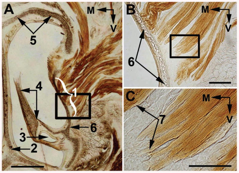
Figure 14.

Light microscopy of an adult rat coronal slice (A) that contains origins (1) of the Partes anterior et interna of the M. nasolabialis profundus. (B) and (C) Higher magnifications of the boxed areas in (A) and (B), respectively. 2, septal cartilage; 3, ductus nasolacrimalis; 4, maxilloturbinate; 5, roof cartilage; 6, lateral nasal cartilage; 7, sites of muscle attachment to the nasal cartilage. M, medial; V, ventral. Scale bars are 1mm in (A), 0.2 mm in (B), and 0.1 mm in (C).
