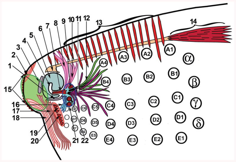
Figure 4.

Schematic representation of muscles related to the dorsorostral part of the rat snout. Encircled areas comprise origins of the following muscles: green area, rhinarial muscles; pink area, two extrinsic whisker protractors; and blue area, most of the parts of the M. nasolabialis profundus. 1, ventromedial part of the lateral nasal cartilage; 2, M. levator rhinarii; 3, septal cartilage; 4, atrioturbinate; 5, atrium; 6, insertion site of the M. dilator nasi; 7, ITM; 8, 10, 11, and 12, the most superficial, superficial, posterior, and pseudointrinsic portions, respectively, of the Pars interna of the M. nasolabialis profundus; 9, origin of the Pars interna profunda; 13, M. transversus nasi; 14, belly of the M. dilator nasi; 15, rhinarium; 16, Pars anterior of the M. nasolabialis profundus; 17, M. depressor septi nasi; 18, M. depressor rhinarii; 19 and 20, origins of the Partes mediae superior et inferior, and 21 and 22, of the Partes maxillares profunda et superficialis, respectively, of the M. nasolabialis profundus. Marked black circles represent mystacial vibrissae.
