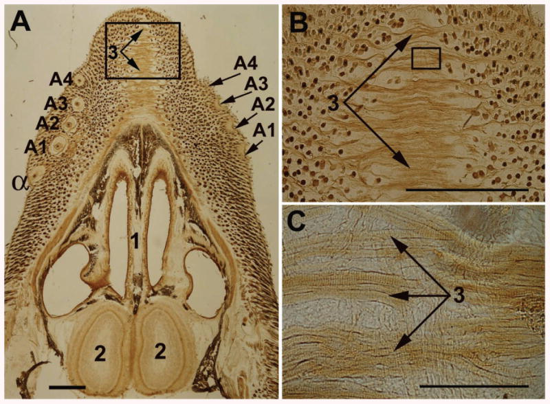
Figure 5.

Light microscopy of a horizontal slice of the dorsal part of a 1-week-old rat snout. (A) A slice of the whole snout; (B) and (C), higher magnifications of the boxed areas in (A) and (B), respectively. α, the dorsal-most straddler; A1 - A4, the dorsal-most row of vibrissae. 1, septum; 2, olfactory bulb; 3, striated fibers of the M. transversus nasi. Scale bars are 1 mm in (A) and (B), and 0.1 mm in (C).
