Figure 3.

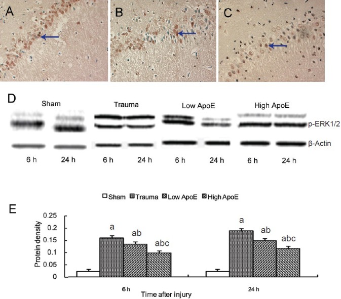
Effects of apolipoprotein E (ApoE) mimetic peptide on extracellular signal-regulated kinase 1/2 (ERK1/2) activation in the hippocampus of rats with diffuse brain injury.
Immunohistochemical staining showed ERK1/2 phosphorylation in neurons in the trauma group (A), low-dose ApoE mimetic peptide group (B) and high-dose ApoE mimetic peptide group (C) at 24 hours after injury (× 200). A few yellow-stained neurons positive for ERK1/2 phosphoryla-tion are visible in the hippocampus in the trauma group, and these neurons are fewer in the low- (low ApoE) and high-dose ApoE mimetic peptide groups (high ApoE). Arrows point to neurons positive for phosphorylated ERK1/2. (D) Phosphorylated ERK1/2 levels were examined by western blot analysis. (E) Bands corresponding to phosphorylated ERK1/2 were scanned and the intensities were normalized to β-actin. Data are expressed as mean ± SD from five rats in each group. Differences between groups were compared with one-way analysis of variance and Student-New-man-Keuls test. aP < 0.05, vs. sham group; bP < 0.05, vs. trauma group; cP < 0.05, vs. low ApoE group. h: Hours.
