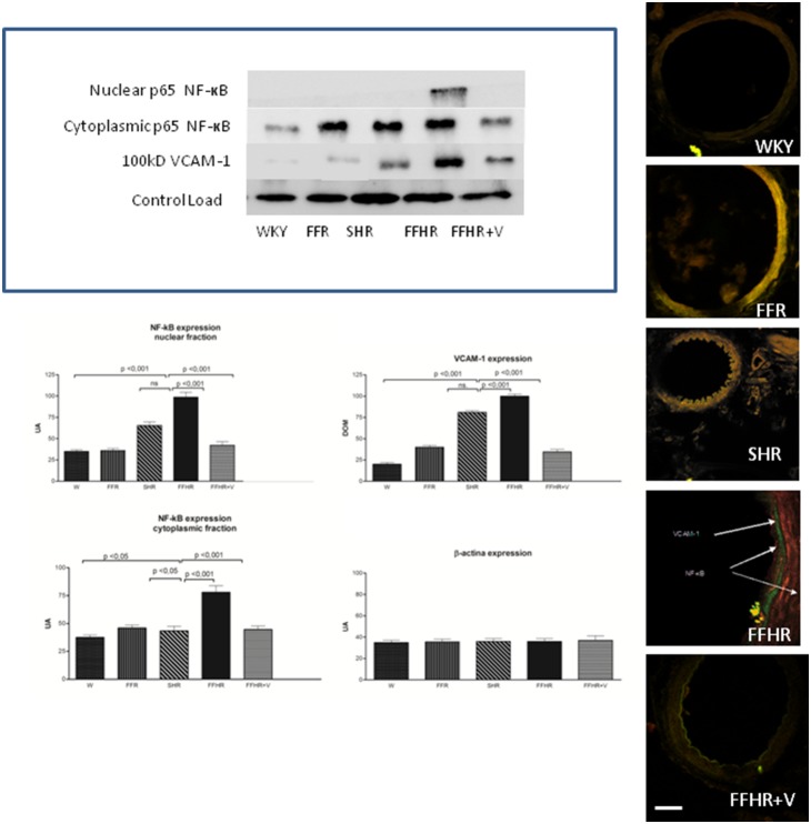
Figure 1. Cytoplasmatic and nuclear p-65 fraction of NF-κB and VCAM-1 expression in mesenteric arteries detected using WB and IHC.
The upper panel shows a representative WB probed with anti-VCAM-1-FITC and anti-p65-TRITC. The results represent the optical density of the bands for each group. The lower panel shows microphotographs of mesenteric tissue obtained using a laser ICM 600x.

