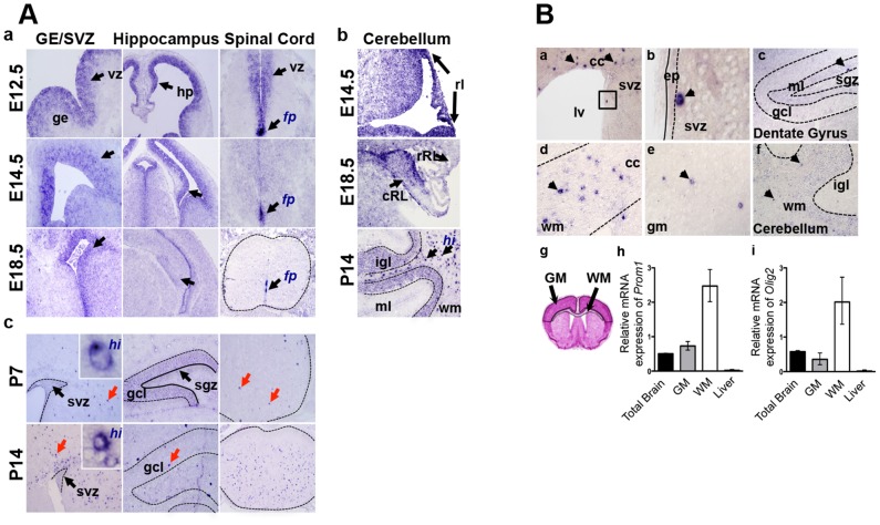
Figure 1. Prom1 expression in developing and adult mouse CNS.
A. RNA in situ hybridization showing high Prom1 expression in the ventricular zone (vz, arrows) of the prenatal brain and spinal cord (a). Levels decrease over time except in the floor plate (fp). In the cerebellum (b) Prom1 is highly expressed in the caudal and rostral rhombic lips (rl, cRL, rRL) while postnatal expression is most intense in rare cells of the white matter (hi, arrows) and the internal granule layer (igl). In postnatal brain (c) expression is conserved at low levels in the stem cell niches (svz and sgz) but is highest in single cells within white matter (wm, red arrows). B. In adult brain Prom1 RNA is highest in single, widely distributed cells (arrowheads) in the corpus callosum (a, d), cerebellum (f), gray matter (e) and only rarely in SVZ (b) and the SGZ (c) stem cell niches. Semi-quantitative RT-PCR of microdissected white and grey matter regions from 3 different mice (g) validate high Prom1 levels in white matter relative to gray matter and whole brain (p = 0.05) (h). Mouse liver acts as a low level reference tissue. The differential level of Prom1 is similar to the oligodendroglial marker Olig2 (i). ge: ganglionic eminence; ml: molecular layer; gm: grey matter; hp: hippocampus; gcl: granule cell layer; sgz: subgranular zone; cc: corpus callosum; lv: lateral ventricle; ep: ependymal layer.

