
Figure 3. Prom1hi cells are slow dividing distributed progenitor cells distinct from adult NG2 progenitors.
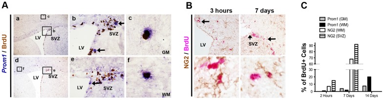
A. Two examples of long term BrdU labeling (7 days, daily dosing) identify rare Prom1+ (RISH; blue) BrdU+ (IHC; brown) cells in white and grey matter (c and f are magnified insets of the small marked area in a and d). High magnification of the SVZ (b, e) shows numerous BrdU+ stem/progenitor cells that do not express high Prom1. Arrows indicate rare Prom1hi+ cells that do not express BrdU (b, e). B. In contrast to Prom1, double IHC for NG2 (brown) and BrdU (red) readily identifies numerous co-localized cells in SVZ and brain parenchyma. C. Manual quantification of the percentage of BrdU positive cells co-labeled with Prom1 or NG2 in the white matter, grey matter and SVZ. svz: subventricular zone; lv: lateral ventricle; gm: grey matter; wm: white matter.