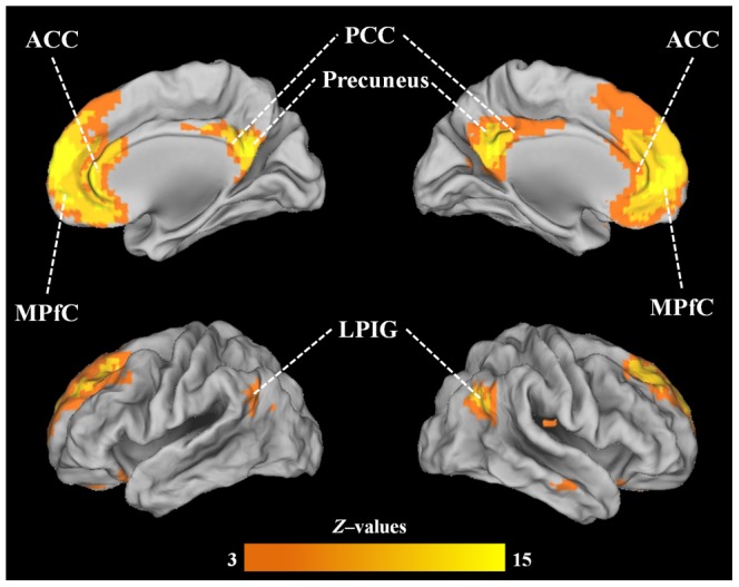
Figure 2. DMN identified in the group analysis of the 20 subjects; the DMN was picked by visual inspection, comprising medial prefrontal cortex (MPfC), anterior and posterior cingulate cortices (ACC and PCC), precuneus and lateral parietal inferior gyri (LPIG).