
Figure 1. The binding pocket of porcine CYP3A29 docked with T-2 toxin.
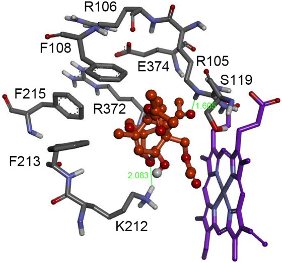
CYP3A29 interaction residues, Arg105, Arg106, Phe108, Ser119, Lys212, Phe213, Phe215, Arg372 and Glu374, are in distances within 5Å to T-2 toxin molecule. The T-2 toxin is colored in orange. The heme is represented in violet color. The green lines and numbers denote hydrogen bonds and bond lengths.