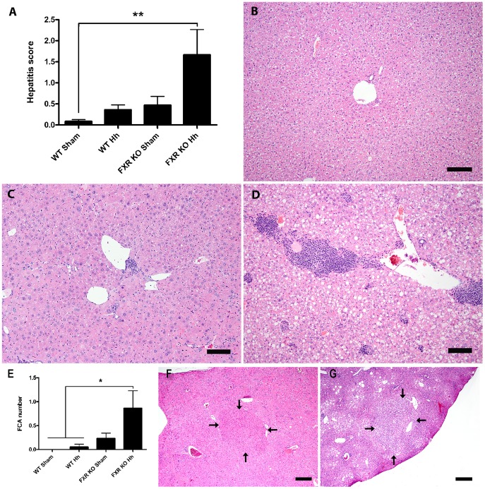
Figure 2. H. hepaticus infection is necessary for increased liver pathology and preneoplastic lesions in FXR KO mice.
A) Mean hepatitis index (HI) ± SD is shown for each experimental group (**P<0.01). Representative liver sections from B) sham-treated WT (HI = 0) and H. hepaticus-infected FXR KO mice showing C) typical (HI = 1.5) and D) severe (HI = 6.5) hepatitis. H&E-stain, bar = 100 µm. E) Mean FCA count ± SD is shown for each experimental group (*P<0.05). Examples of F) eosinophilic and G) clear cell foci from H. hepaticus-infected FXR KO mice (outlined by arrows; H&E-stain, bar = 600 µm).

