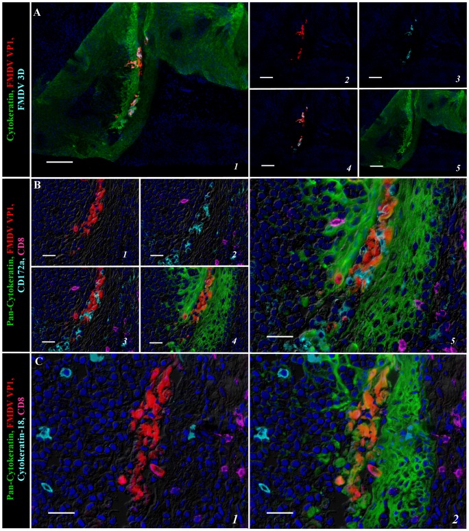
Figure 3. Multichannel immunofluorescent detection of FMDV structural (VP1) and non-structural (3D) protein in porcine paraepiglottic tonsils at 24 hpi.
A) FMDV VP1 (red) and 3D (turquoise) proteins co-localize with cytokeratin (green) in regionally expanding foci of primary FMDV infection within reticular crypt epithelium of the paraepiglottic tonsil. 10× magnification, scale bar 100 µm. B) Serial section of region identified in (A). Localization of FMDV VP1(red) is restricted to cytokeratin-positive epithelial cells (green); leukocytes expressing CD172a (turquoise) and CD8 (purple; presumptive NK cells) are interspersed amongst virus-infected cells. 40× magnification, scale bar 50 µm. C) Serial section of region identified in (A–B). Cytokeratin-18 expressing M-cells (turquoise) localized within segments of epithelium containing FMDV VP1-positive cells (red), but without co-localization. 40× magnification, scale bar 50 µm.

