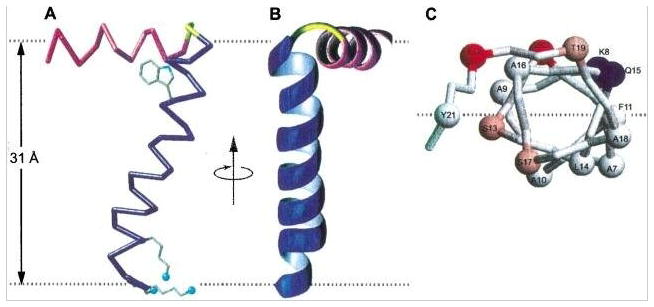
Figure 1.

Three views of the structure of the membrane-bound form of fd coat protein in POPC/POPG phospholipid bilayers. The amphipathic in-plane helix is in magenta, the hydrophobic trans-membrane helix is in blue, and the short connecting turn is in yellow. The flexible N- and C- terminal residues are not shown. A. Side view of TM helix, the Trp and Lys sidechains are shown in blue. The dashed gray lines mark the lipid-water boundary. B. Front view and, C. View of in-plane helix looking down from the C-terminus. From Marassi & Opella (2003).
