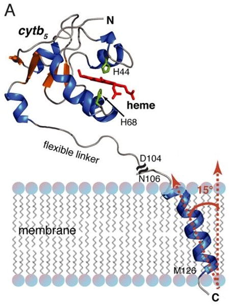
Figure 6.

NMR structure of rabbit microsomal cytb5. NMR structure of full-length cytb5 obtained from a combined solution and solid-state NMR approach. The soluble heme domain structure (residues 1–104) of full-length cytb5 was solved in DPC micelles by solution NMR. The transmembrane domain structure (residues 106–126) of full-length cytb5 was determined in aligned DMPC/DHPC bicelles using solid-state NMR spectroscopy. From Ahuja et al (2013).
