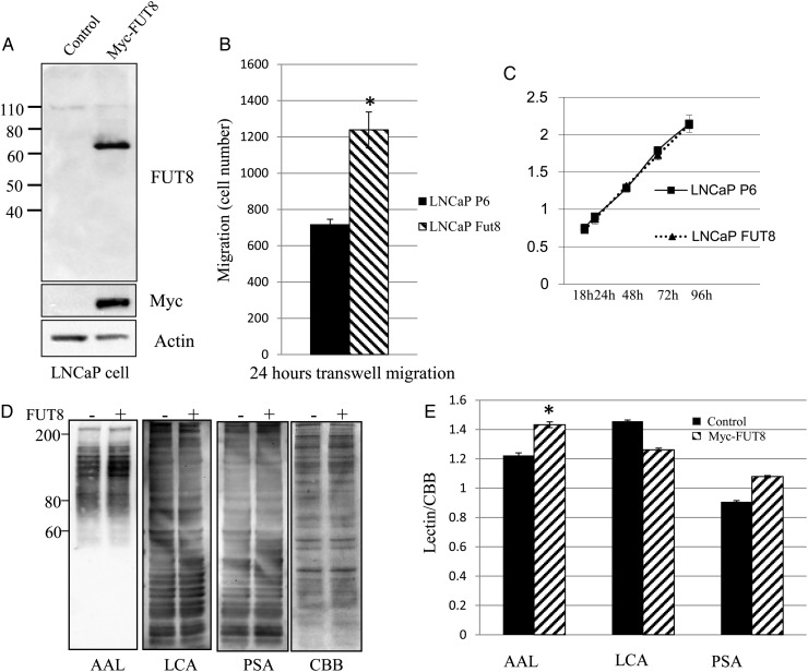
Fig. 6.
FUT8 overexpression increases LNCaP cell motility. (A) LNCaP cells stably expressed Myc-FUT8. LNCaP cells were transfected with empty vector control P6 (Control P6) or Myc-FUT8. After screening and expanding, whole cell lysates were subjected to western blot using anti-FUT8 and anti-Myc monoclonal antibody. (B) Transwell migration assay of LNCaP cells transfected with either empty vector control P6 (LNCaP P6) or Myc-FUT8 (LNCaP FUT8). Cell numbers at the bottom of transwell chamber were counted 24 h after migration. * P < 0.05, data represented mean ± SD (n = 3). (C) Cell proliferation was determined by MTT assay using Cell Counting Kit-8. LNCaP cells were transfected with either empty vector control P6 (LNCaP P6) or Myc-FUT8 (LNCaP FUT8). Data represented mean ± SD (n = 3). (D) Whole cell lysates from either empty vector control P6 (P6) or Myc-FUT8 transfected LNCaP cells were subjected to lectin blot analysis using AAL, LCA and Arachis hypogaea lectin (PSA). CBB staining of gels shows comparable amounts proteins in each lane. (−) empty vector control; (+) Myc-FUT8. (E) Optical density of lectins staining was quantified by CBB staining using NIH ImageJ. In AAL lectin staining result, the P-value is <0.05 (*).

