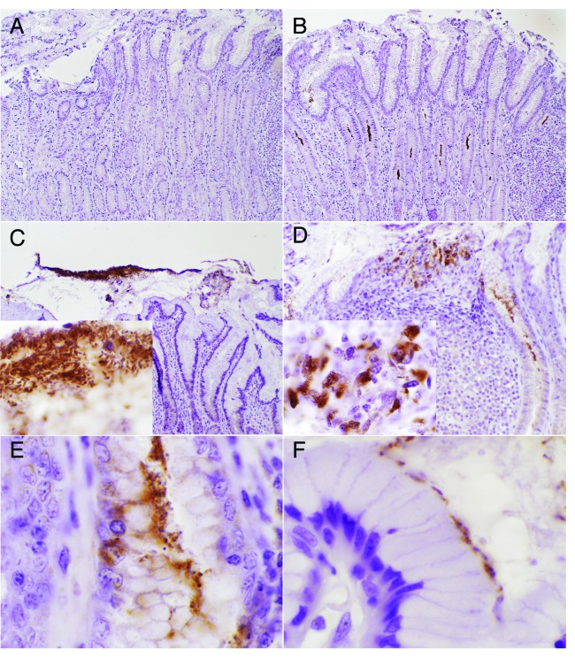
Figure 1. Localization of Helicobacter pylori strain SS1 in the mucosa of the pig stomach at day 50 post-infection. Bacterial detection was performed in formalin fixed stomach sections stained with an anti-H. pylori polyclonal antibody from Cell Marqe, or with secondary only as a negative control (A). Most of the H. pylori was localized in the mucus layer and gastric pits (B, C), however, a small fraction was found in the lamina propria (E). At high power magnification (1000×) H. pylori can be observed in the extracellular compartment as free swimming (C, insert) or overlying the apical side of epithelial cells (E). Some H. pylori positive staining was detected in the intracellular compartment of cells in lymphoid aggregates (D, insert) and within epithelial cells (E). Original magnification, 100× (A, B), 200× (C, D), and 1000× (C, insert; D, insert; E and F).

An official website of the United States government
Here's how you know
Official websites use .gov
A
.gov website belongs to an official
government organization in the United States.
Secure .gov websites use HTTPS
A lock (
) or https:// means you've safely
connected to the .gov website. Share sensitive
information only on official, secure websites.
