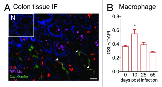
Figure 2. Macrophage recruitment and RELMα expression in C. rodentium-infected mice. (A) Immunofluorescent stained colon tissue sections from naïve (N) or day 10-infected WT mice reveal GSL+ macrophages (red), RELMα (purple), C. rodentium-GFP (green), and DAPI (blue). Scale bar 25 μm. (B) Quantification of GSL+ macrophage frequency in naïve or infected mice was performed.
