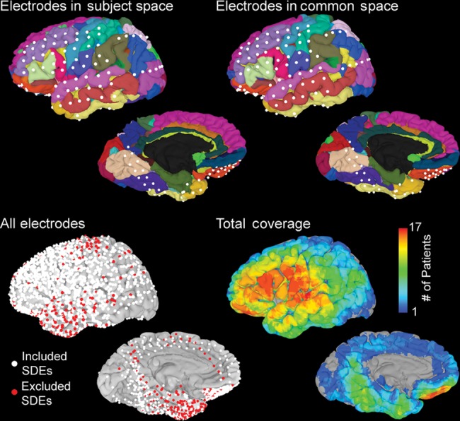
Figure 2.

Distribution of electrodes used in the analysis. (Top, left) SDEs localized onto individual subject space and viewed on an automatically parcellated cortical surface. (Right) Using a rigid, 12-parameter affine transformation, electrodes were aligned with the MNI-N27 brain in the Talairach coordinate space. (Bottom) All electrodes for all subjects transformed into the MNI-N27 space and displayed on the surface. SDEs over epileptogenic tissue or those with significant noise (red, n = 313) were removed from the analysis. The remaining electrodes (white, n = 1629) were used in the group analysis and to generate a total coverage map.
