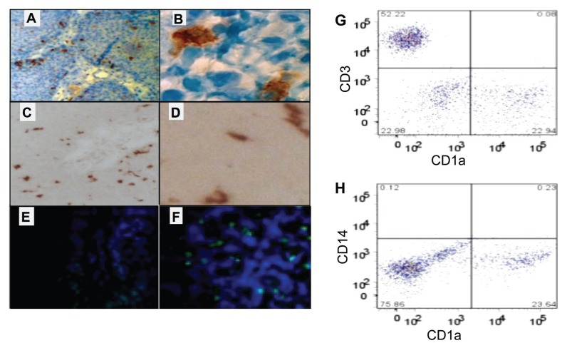
Figure 3.
Papillomas contain abundant iLCs. iLCs were CD1a+ (A,B) and langerin+ (C,D), but CD83− (E). A positive control (lymphoma) was CD83+ (F). (A–B) Paraffin sections, DAB stain. (C–D) frozen sections, AEC stain. (E–F) Paraffin sections, immunofluorescence with DAPI counterstain. (G, H) Flow cytometry of LCs isolated from papilloma biopsies. LCs (G, lower right) were approximately half as frequent as T-cells (G, upper left). LCs were CD14 negative (H, lower right), confirming that they were not monocytes and that there was no significant contamination by blood.

