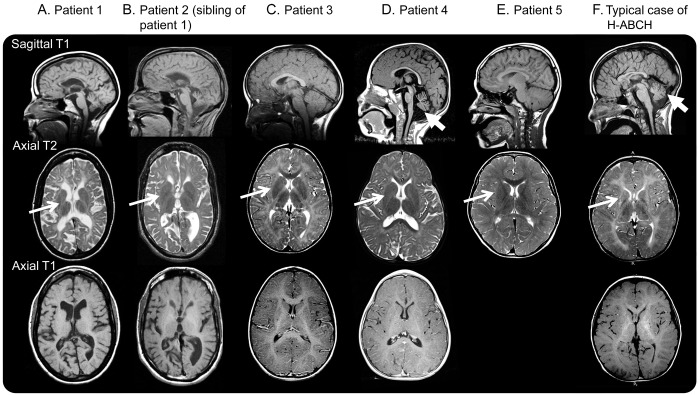
Figure 1. Brain sagittal T1, axial T2, and axial T1 MRIs of participants.
(A) Patient 1 at age 45 years. Note the generalized atrophy with preserved putamen (white arrow) and hypomyelination (T1- and T2-hyperintensity of the cerebral white matter). (B) Patient 2 at age 42 years. Note the generalized atrophy with preserved putamen (white arrow) and hypomyelination (T1- and T2-hyperintensity of the cerebral white matter). (C) Patient 3 at age 4 years. Note the hypomyelination (T1- and T2-hyperintensity of the white matter) with preserved putamen (white arrow) and cerebellum. Only postcontrast T1-weighted image available. (D) Patient 4 at age 5 years. Note the preserved putamen (white arrow) and hypomyelination (T1- and T2-hyperintensity of the white matter). Mild atrophy of the cerebellar vermis is seen (thick white arrow). Only postcontrast T1-weighted image available. (E) Patient 5 at age 10 years notable for preserved putamen (white arrow) and mild hypomyelination (T1- and T2-hyperintensity of the white matter). No axial T1-weighted images were available. A repeat study at 14 years showed no putamen atrophy. (F) Typical H-ABC MRI of the brain at age 9 years with absences of putamen (white arrow) and hypomyelination (T1- and T2-hyperintensity of the white matter), as well as cerebellar atrophy (thick white arrow). H-ABC = hypomyelination with atrophy of the basal ganglia and cerebellum.

