
Fig. 4.
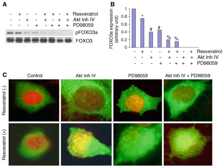
Inhibition of FOXO3a phosphorylation by inhibitors of AKT, MEK/ERK, and resveratrol. a HUVEC cells were pretreated with AKT inhibitor IV (1 μM) and/or MEK1/2 inhibitor PD98059 (10 μM) for 2 h, followed by treatment with or without resveratrol (20 μM) for 24 h. Cells were harvested and cytoplasmic fractions were prepared. Crude proteins were subjected to Western blot analysis to measure the expression of phospho-FOXO3a and total FOXO3a. b Expression density of pFOXO3a. The expression density of phospho-FOXO3a protein was quantified from Western blot reported in (a), and plotted on Y-axis. The Control band was normalized to 1. *, #, and % significantly different from respective control, P < 0.05. c Nuclear translocation of FOXO3a. HUVECs were seeded in chambered slides and treated with or without resveratrol (20 μM) for 18 h. Cells were fixed, permeabilized, and stained with anti-FOXO3a antibody at 4°C for 18 h. After washing, cells were stained with DAPI (nuclear staining) and secondary antibody conjugated with FITC, and visualized under a fluorescence microscope. Green color FOXO3a, red color nucleus (for clarity the color of nucleus was changed from blue to red), yellow colocalization of FOXO3a to nucleus (Color figure online)