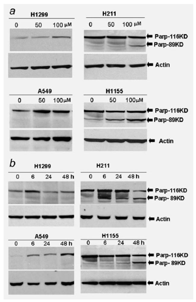
Figure 2.

Western immunoblot analysis of PARP cleavage induced by PRIMA-1 in lung cancer cell lines. (a) Human lung cancer cells A549 (wild-type p53), H1299 (p53 null), H211 (mutant p53, 248Q) and H1155 (mutant p53, 273H) were treated with PRIMA-1 at a dose of 50 or 100 μM. Cells were harvested 24 hr posttreatment. A total of 100 μg cell lysate each sample was subjected for Western immunoblot analysis. Western immunoblot membrane was probed with antibodies against full length and cleaved PARP. (b) Human lung cancer cells were treated with PRIMA-1 (100 μM). Cells were harvested and lysed at 6, 24 and 48 hr posttreatment. Western immunoblot analysis was used to analyze the cleavage of PARP protein in the lung cancer cells at indicated time point. Membrane was probed with antibodies against full length and cleaved PARP. Loading control was β-actin. The cleaved PARP was detected in the H211 and H1155 cells after treatment with PRIMA-1.
