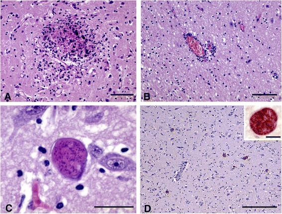
Figure 1.

Histological findings in foetus/lamb brains positive to Neopora caninum PCR. A) Focus of gliosis with central area of coagulative to caseous necrosis in the grey matter (HE; Bar = 100 μm); B) Mild perivascular infiltration of mononuclear inflammatory cells in the white matter (HE; Bar = 200 μm); C) Intracellular cyst containing high number of bradyzoites. Note the absence of inflammation (HE; Bar = 25 μm); D) Immunohistochemical labelling of several protozoan tissue cysts in the grey matter. Note the absence of evident inflammatory infiltration in relation to the cysts (IHC; Bar = 300 μm) Inset: Detail of an immunolabelled tissue cyst (IHC; Bar = 15 μm).
