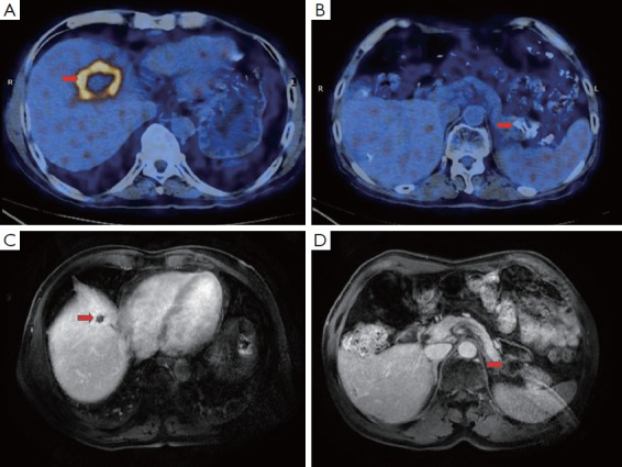
Figure 1.

Imaging evaluation of the primary and metastatic lesions before and after treatment. (A,B) PET/CT scanning revealed a 57 mm × 37 mm soft mass in the pancreatic tail and multiple nodules in the liver (sized from 12 mm × 12 mm to 54 mm × 37 mm); (C,D) MRI performed in February 2013 demonstrated a favorable prognosis for the patient, indicating a decreased size for the primary tumor at the pancreatic tail (20 mm × 20 mm) for and the hepatic lesions (sized 12 mm × 12 mm to 30 mm × 20 mm).
