
Figure 1.
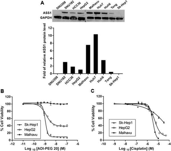
Effect of ASS1 expression on the sensitivity of HCC cell lines to ADI-PEG 20 and cisplatin treatment. (A) ASS1 protein levels from cell lysates were determined by western blot. GAPDH was run as a loading control. The signal intensities of the bands were quantified and normalized by taking the level of HepG2 cells as 1. (B and C) Sensitivity of three representative HCC cell lines to ADI-PEG 20 and cisplatin. Cells were cultured in medium containing various concentrations of (B) ADI-PEG 20 and (C) cisplatin. Duplicate samples were assessed for cell viability after 72 h using the Promega luminescence assay. Percent cell viability from either ADI-PEG 20 or cisplatin-treated cells was calculated relative to the viability in corresponding matched DMSO-treated cells, which was designated as 100%. The data are representative of three or more independent experiments. Error bars represent S.D.