Figure 1.

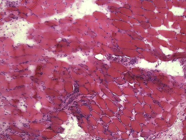
H&E-stained section (×10 magnification) reveals moderate infiltration by lymphocytes (confirmed by immunohistochemistry for CD45 and CD3 (not shown). Frequent severely atrophic fibres are present, often showing signs of regeneration.

H&E-stained section (×10 magnification) reveals moderate infiltration by lymphocytes (confirmed by immunohistochemistry for CD45 and CD3 (not shown). Frequent severely atrophic fibres are present, often showing signs of regeneration.