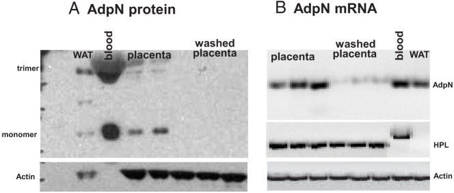
Figure 2.

Effect of blood removal on adpN expression in the placenta. A) representative Western blot showing detection from tissues of normal-weight women at term delivery. The adpN monomer migrated as a 33-kDa apparent molecular weight signal, and the dimer migrated as a 60-kDa signal. Identical results were obtained from 4–6 independent experiments. B) RT-PCR analysis of adpN mRNA. Total RNA was extracted from human placenta before and after blood removal through serial PBS washes and was analyzed by RT-PCR. Maternal adipose tissue and blood were used as positive controls. The number of amplification cycles was 45 for adpN and 30 for β-actin. A nonspecific HPL amplification product was detected in white blood cells. Identical results were obtained from 4–6 independent experiments. WAT, white adipose tissue; bl, blood.
