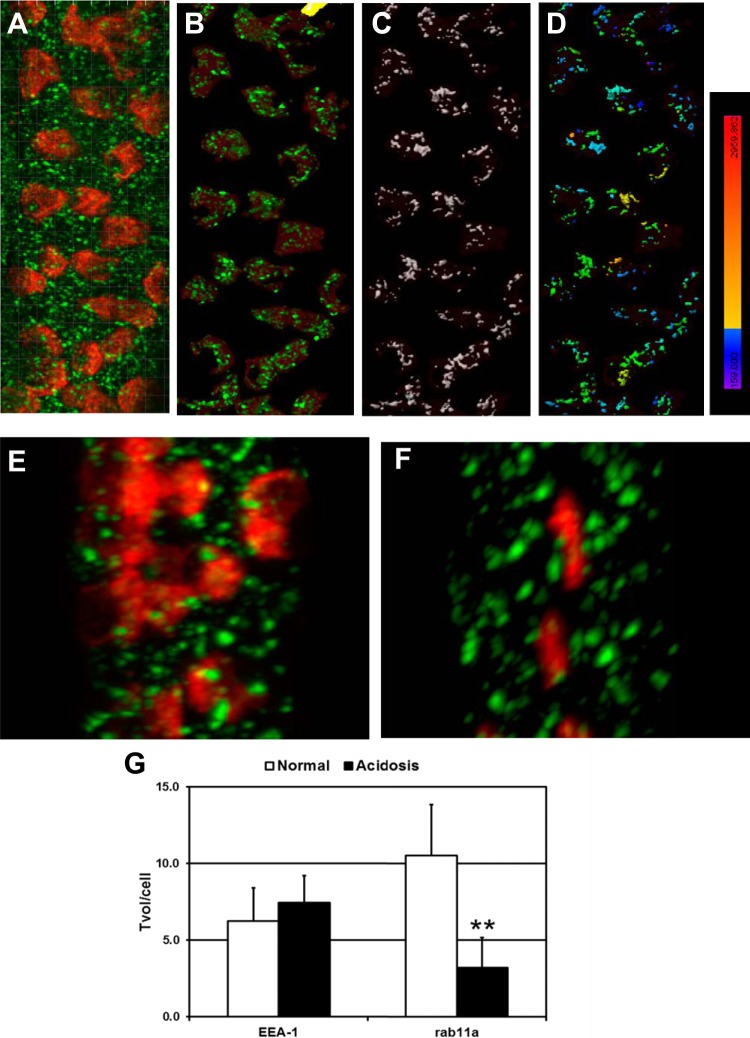
Fig. 3.
Acidosis reduces the volume of the Rab11a+ apical recycling endosome compartment in β-ICs. A: 3-D projection of a microdissected CCD from normal rabbit kidney stained for pendrin (red) and early endosomal antigen (EEA)-1 (green). Note that colocalization algorithms were not used to identify pixel overlap between red and green stains in this image. B: identification of pendrin cap surfaces with Imaris software enabled the exclusion of EEA-1-labeled vesicles residing outside pendrin caps. C: demarcation of EEA-1 surfaces within pendrin cap boundaries. D: chromatic scale illustrating the intensity of pendrin staining with spatial overlap with EEA-1-labeled surfaces. The numeric values associated with the chromatic scale ranged from 159 (purple) to 2,960 (bright red). E and F: microdissected CCDs from normal (E) or acidotic (F) rabbit kidneys stained for pendrin (red) and Rab11a (green). G: total vesicular volume in β-ICs (Tvol/cell) for EEA-1+ and Rab11a+ compartments. Results are from up to 5 rabbits (n = 3–5), 9–17 CCDs, and 283–659 β-IC per acid-base condition. **The normal → acidosis Rab11a total volume per cell number is different at the 2% confidence interval (by Mann-Whitney U-Test).

