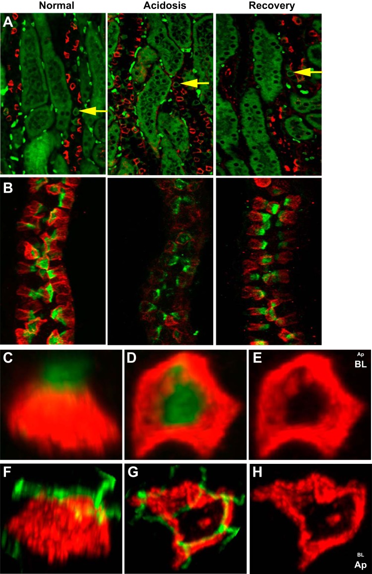
Fig. 4.
AE4 (SLC4A9) is expressed as a “barrel-like structure” localized below ZO-1 in β-ICs. A: kidney sections prepared from rabbits with the indicated acid-base conditions were stained for AE4 (red) and AE1 (green) and photographed as described in materials and methods. Yellow arrows point to representative AE1-positive cells located in CCDs. The intensely stained green dots in the interstitium are red blood cells. B: microdissected CCDs isolated from rabbits with the indicated acid-base conditions and stained with peanut agglutinin (PNA; green) and antibody directed against AE4 (red). Z-stack images were obtained by confocal microscopy. C–E: 3-D reconstructions of an individual β-IC from a normal rabbit CCD stained as in B. C: lateral view of a β-IC. D: 90° rotation from the lateral view such that the PNA cap is facing the background and the basolateral surface is in the foreground. E: AE4 distribution sans the PNA cap. F–H: individual β-ICs from a normal rabbit CCD stained for AE4 (red) and ZO-1 (green). F: lateral view of a β-IC. G: 90° rotation bringing ZO-1 and the apical surface to the foreground. H: AE4 distribution sans ZO-1.

