
Figure 1. Expression of mouse Mbd5 transcripts.
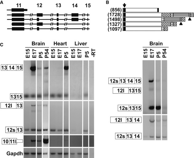
A Schematic of the Mbd5 transcripts depicting exons 11–15.
B Schematic of the predicted mouse MBD5 proteins. The black box (arrow) represents the MBD, the gray box (arrowheads) the PWWP domain, and the shaded areas represent alternative regions of the proteins. In brackets is the number of amino acids.
C RT–PCR from cDNA derived from mouse brain, heart, and liver at the indicated developmental time points. The numbered boxes represent the exons included in the amplicons. 12l and 12s depict the extended and short exon 12, respectively; and 11l depicts the extended exon 11. The data show that 11l is a terminal exon since we did not detect transcripts carrying exon 11l spliced with any downstream exon.