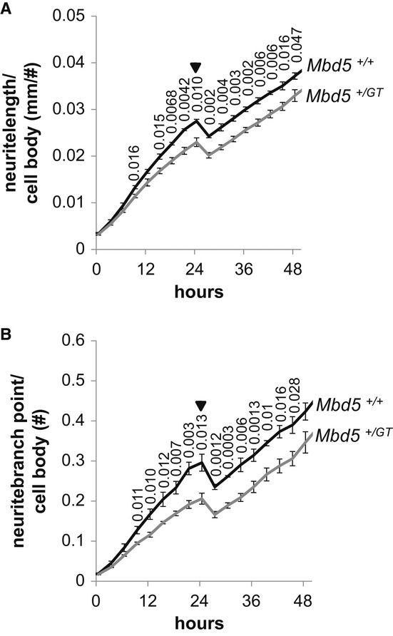
Figure 7. Mbd5+/GT neurons have reduced neurite outgrowth and branching.

A, B Dissociated E16 cortical neuron cultures generated from Mbd5+/GT and Mbd5+/+ mice were plated, and cellular morphology was recorded every 3 h for 48 h using an IncuCyte live-cell imaging system located within the incubator. Phase images were analyzed for process length and branching by Incucyte's NeuroTrack software. Shown are average values for neurite length (A) and number of branching points (B) normalized to cell bodies of quadruplicate wells per embryo (n = 3 Mbd5+/+ and n = 4 Mbd5+/GT; Student's t-test; unpaired, two-tailed distribution, P-values are displayed above the line in the figure). Arrows indicate the removal of the plates from the incubator at 24 h to replace plating media for neurobasal media.
