
Figure 8. Mbd5 acts in vitro as a transcriptional activator.
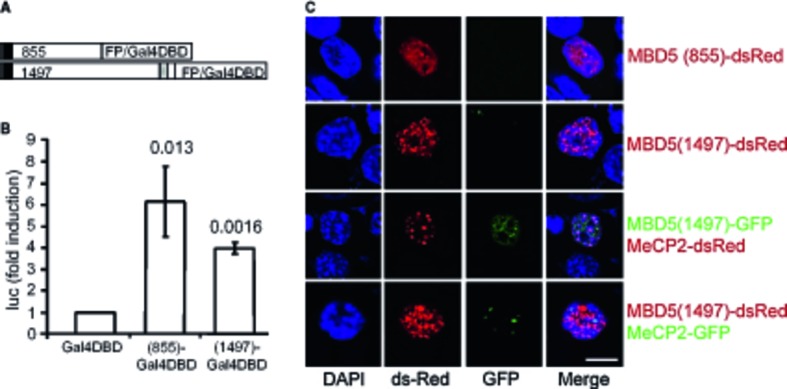
A Schematic of the constructs for the transactivation studies. The black box represents the MBD, and the gray box the PWWP domain.
B Constructs encoding the Gal4 DNA-binding domain fused to Mbd5 and the Gal4-responsive luciferase reporter gene were co-transfected into HEK293 cells. A β-galactosidase expression plasmid was also co-transfected to allow normalization for transfection efficiency. Cells were collected, and luciferase activity was measured 24 h after transfection. Each bar represents the mean ± SD from three independent experiments. (Student's t-test; unpaired, two-tailed distribution compared with cells transfected with GAL4-DBD, P-values are displayed above the bars in the figure.)
C Localization of transfected Mbd5 in mouse neuroblastoma N2A cells. Mbd5 and the heterochromatic marker MeCP2 fused to GFP or dsRed at the N-terminus were transfected into N2A cells. DAPI was used for nucleic acid staining (blue). The images were taken with a confocal LSM 710 microscope from Zeiss. Scale bar = 10 μm.