Figure 2.

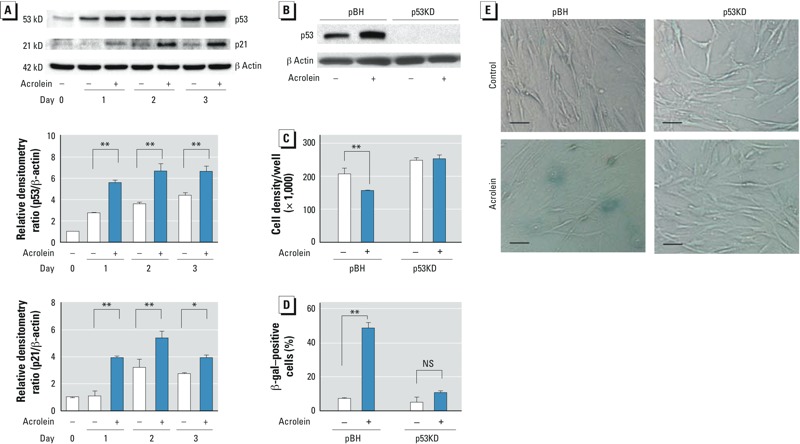
Effects of siRNA-mediated p53 suppression on cellular senescence. (A) p53 and p21 proteins in HFL‑1 cells cultured in the absence (–) or presence (+) of 25 μM acrolein for 1, 2, or 3 days. Immunoblot analysis of p53 and p21 (top), and the relative densitometry ratio for p53/β‑actin (center) and p21/β‑actin (bottom); each day 0 control was regarded as 1.0. (B) Immunoblot analysis of transduced HFL‑1 cells [p53KD (p53-deficient) and pBH (control)] cultured in the presence or absence of 25 μM acrolein for 2 days; the blot is representative of three experiments. (C–E) p53KD and pBH cells were cultured for 3 days with or without 25 μM acrolein and then subcultured (at a starting density of 50,000 cells/well) for 3 days. (C) Cell density of subcultured p53KD and pBH cells. (D) Percentage of SA β-gal–positive cells per total cell number 3 days after subculture. (E) Representative photomicrographs of p53KD and pBH cells 3 days after subculture (bars = 5 μm. In A, C, and D, data are expressed as mean ± SEM of three independent experiments. NS, not significant. *p < 0.05. **p < 0.01.
