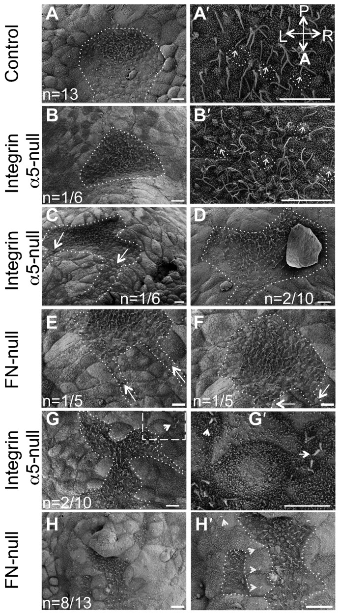
Fig. 1. Integrin α5 and fibronectin are required for morphogenesis of the mouse node.

(A,A′) Shape of the node in the control mouse embryo at E7.5. (A) The dotted line outlines the entire shape of the node. (A′) Primary cilia (arrows) are positioned at the posterior of nodal cells (some examples are outlined). (B,B′,C,D,G,G′) Node shapes are aberrant in integrin α5-null E7.5 embryos. (B) Notice the inverted shape of the node in this integrin α5-null mutant. Nodes were inverted in 6 out of 10 integrin α5-null mutants. (B,C) Examples of such inverted nodes. (B′) Magnified view of cilia in the node shown in panel B. Each primary cilium (arrows) is positioned at the posterior of each cell (outlined). (C) Note two notochords emanating from each corner of a mutant node (dotted line, arrows). (D) The pointy end of the node was positioned in a correct orientation in 2/10 integrin α5-null mutant nodes (dotted line outlines the node and the notochord). (E,F,H,H′) Aberrant nodes in E7.5 FN1-null mutants. (E,F) Two examples of inverted nodes in FN1-null mutants. Nodes were inverted in 5 out of 13 FN1-null mutants. (G,H) In some α5-null and FN1-null mutants, the nodes were narrow and disrupted. (G′,H′) Magnified views of panels G and H. Areas containing node cells are outlined. Arrows point to cilia protruding from beneath the cells of the primitive endoderm. A–anterior, P–posterior, L–left and R–right axes are marked and their directions are the same in all panels. Scale bars: 10 µm.
