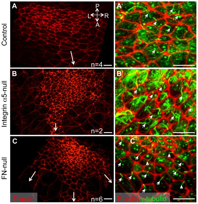
Fig. 2. Anterior–posterior polarity of the nodal cells does not depend on integrin α5β1 or FN1.

Red: F-actin is detected using rhodamine-phalloidin; Green: acetylated (stable) α-tubulin. Positions of the notochords are marked by long arrows in panels A–C. Note three notochords originate from the middle and the two corners of the inverted node in this FN1-null embryo (arrows, C). Protruding cilia (small arrows, A′–C′) are located at the posterior of each nodal cell. Axes are marked as in Fig. 1. All embryos were collected in the morning of E8.0. Scale bars: 10 µm.
